 AI Assistant

Three-photon microscopy is an advanced nonlinear optical imaging technique that uses ultra-short pulsed infrared lasers as the light source. Due to the strong penetration ability of infrared light, three-photon imaging can penetrate deep into tissues without the significant scattering or absorption that occurs with visible light at the surface. This gives the technique a significant advantage in high-resolution imaging of deeper tissues. Compared to traditional single-photon and two-photon microscopy, three-photon imaging provides greater imaging depth and clearer cellular or tissue structure information, making it highly suitable for applications in neuroscience and biomedical research.
Currently, the exploration of the brain mainly relies on optical microscopy, but the scattering of light by neural tissue prevents the light beam from penetrating deeply into the brain.
The insufficient imaging depth significantly hinders the quality of brain neural imaging.Therefore, achieving deep brain imaging has become an important research focus in brain neural imaging.
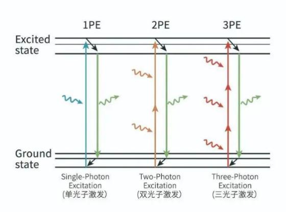
The emergence of multiphoton microscopy has alleviated, to some extent, the challenge of insufficient imaging depth. Multiphoton imaging is based on the multiphoton excitation effect. Fluorescent molecules have discrete energy levels, with the lowest energy level called the ground state, where most molecules reside, and the higher energy levels called the excited states. When illuminated with light of a specific wavelength, electrons in the ground state can absorb multiple photons and transition to the excited state. After remaining in the excited state for a certain period, the electrons relax and transition back to the ground state, emitting a photon with a corresponding wavelength. If the incident light wavelength is longer, it may cause the ground state molecules to absorb multiple photons simultaneously, leading to multiphoton excitation.
Three-photon vs Two-photon
|
|
|
|
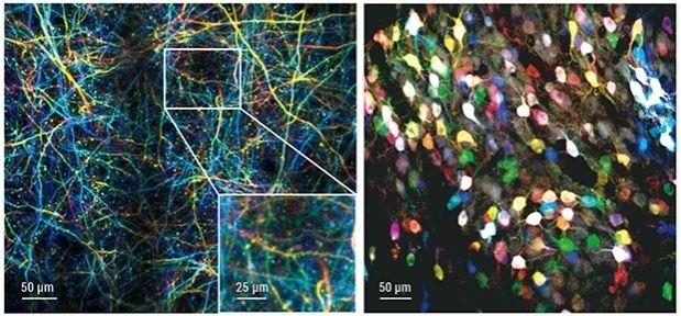 |
Single dendritic spine-level resolution under a 640 µm field of view. Thy1-YFPH transgenic mouse (left) and wild-type mouse brain cortex injected with AAV-hSyn-GCaMP6s virus (right). Probe model: FHIRM-U. Imaging depth: 0-60 µm projection (left), 200-260 µm projection (right). Excitation wavelength: 920 nm. Imaging of freely moving mouse. Right image: Miniature three-photon microscope records structural and functional dynamics in the mouse brain cortex (L1-L6) and hippocampus CA1. |
|
 |
|
CC: Corpus callosum. Green represents GCaMP6s-labeled neuronal calcium signals, while magenta represents third-harmonic signals from the dura mater, microvasculature, and white matter interface. |
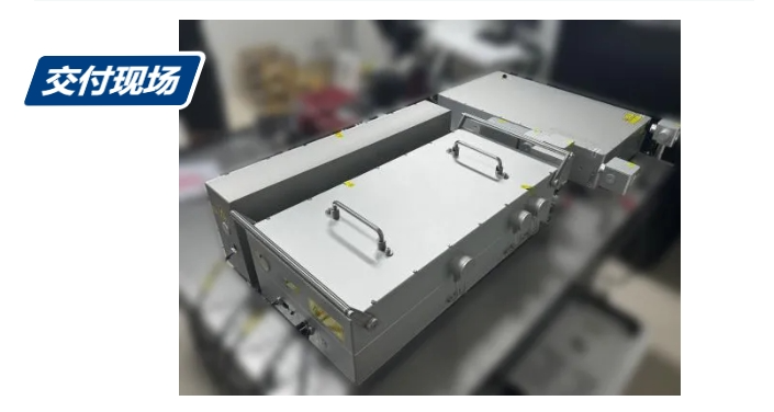
On-site delivery:
|
|
|
Specification:

1)Lower repetition rates and higher pulse energies are available upon request.
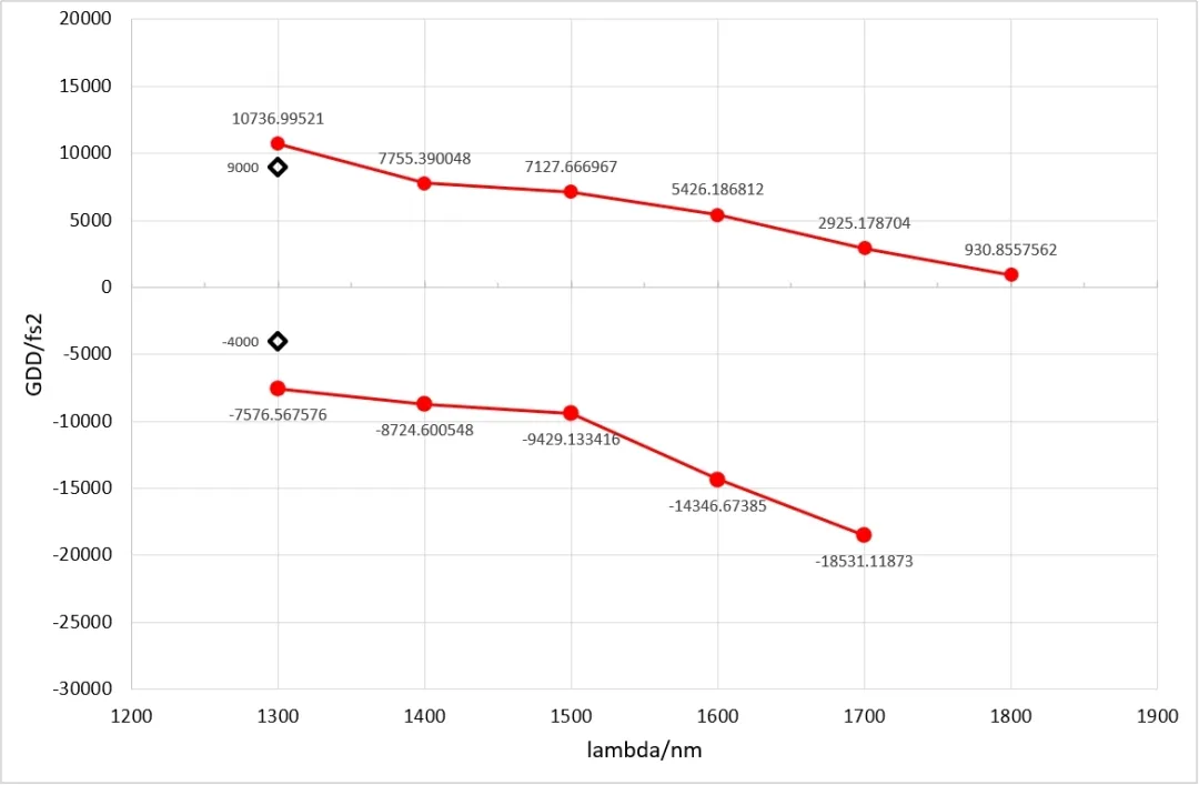
2)Continuous dispersion control; −3000 fs² to +3000 fs² compensation for microscopy.
3)Pump laser: 50W / 50µJ / 1030nm, supports higher power customization. Please contact us for details.
4)Measured after compression at 1/e²
5)Optional expansion for 650-900nm output available. Please contact us for details.
Measured parameters:
|
|
|
|
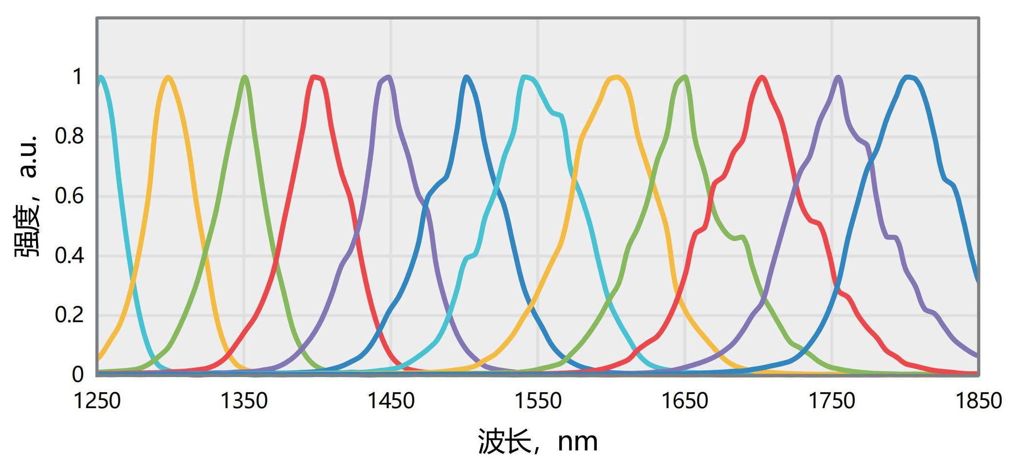
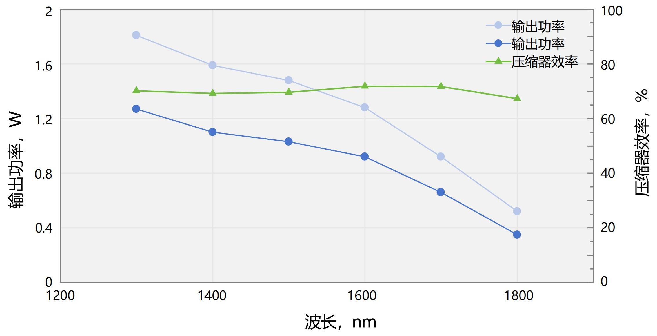
| Typical spectrum (1250-1800nm) | Three-photon system + compressor output power comparison (drive laser :1MHz/40uJ/301fs@HELIOS-40W) |
 |
|
|
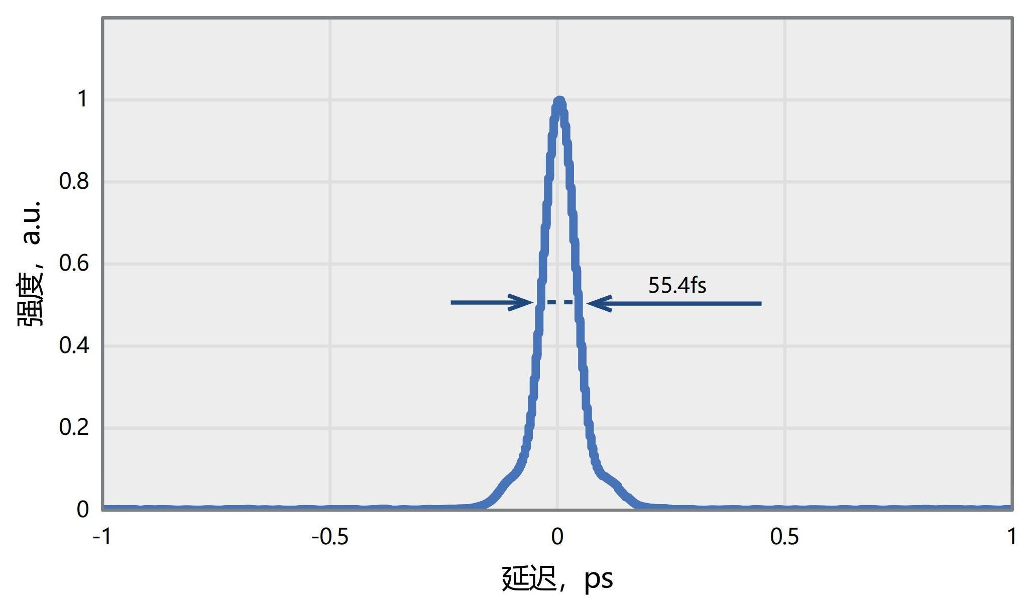
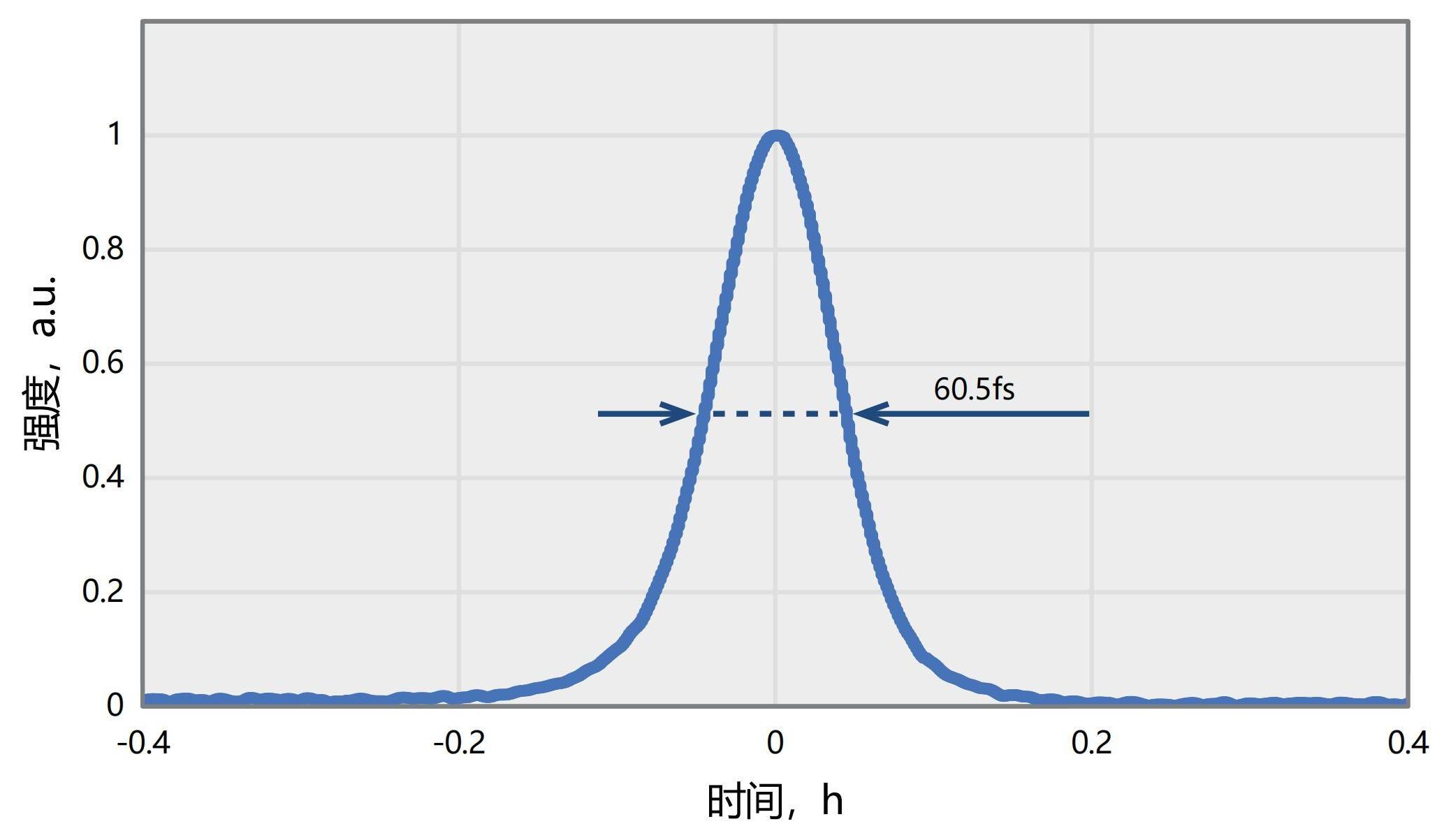
| Typical pulse width(1300nm@55.4fs) | Typical pulse width(1700nm@60.5fs) |
 |
|
|
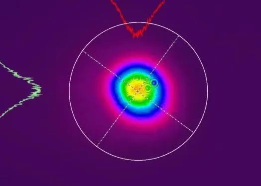
| Output spot @1300nm (compressor outlet 500mm) | GDD Rang |
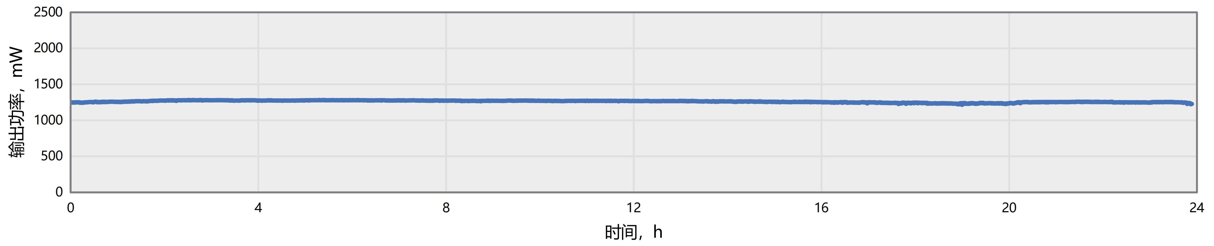
AURORA-3P series 24H output power stability RMS = 0.4289% @ 1300nm
In vivo imaging for biological imaging applications:
|
|
||
|
|
|