 AI Assistant
With femtosecond laser probe drilling technology, doctors can precisely control the cutting process, obtaining the required tissue samples with accuracy while avoiding damage to adjacent healthy tissue. This technology is particularly important in tumor biopsy, where it can be used to accurately cut tumor tissue to obtain samples for pathological analysis. This is especially useful for tumors that are difficult to reach or are located in sensitive areas, such as brain tumors. Furthermore, in the study of heart disease, femtosecond laser technology also demonstrates its value by allowing for the precise collection of small heart tissue samples without damaging the surrounding myocardial tissue.
BIOPSY PROBE DRILLING
2023.11.30
Biopsy probe drilling technology is an important medical surgical technique that allows doctors to extract samples from living tissue for pathological examination and diagnosis. This technology is especially crucial in fields such as oncology and pathology, as it enables doctors to obtain accurate diagnoses of diseases, playing a key role in cancer diagnosis and treatment decisions. The biopsy technique involves using a special probe to penetrate tissue and collect samples. This method allows doctors to obtain samples from specific, sometimes hard-to-reach areas of tissue, while minimizing damage to the surrounding healthy tissue.
The introduction of femtosecond laser technology has further enhanced the precision and safety of biopsy probe drilling. Compared to traditional laser technologies, such as nanosecond lasers, femtosecond lasers, with their extremely short pulse durations, significantly reduce thermal damage during tissue cutting, thereby improving the precision and safety of the procedure. The high precision and minimally invasive nature of this technology make it a vital tool in medical surgery, especially in situations where precise control over cutting depth and location is required.
The ultrashort pulses generated by femtosecond lasers not only allow for highly precise cutting in tissue, but also significantly reduce the risk of thermal damage and inflammation due to localized energy deposition. This contactless cutting method, compared to traditional mechanical drilling techniques, reduces tissue compression and damage, while the short pulse duration helps minimize heat conduction and reduces thermal damage to surrounding tissues. This makes the biopsy process more minimally invasive, reducing surgical trauma and recovery time for patients.
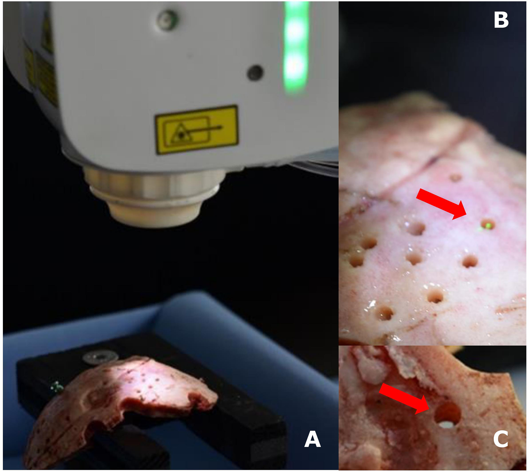
The experimental setup consists of a navigated Er: YAG laser and a skull (A), a detailed photo of the laser in operation (B), and the hole formed by the laser on the skull (C).
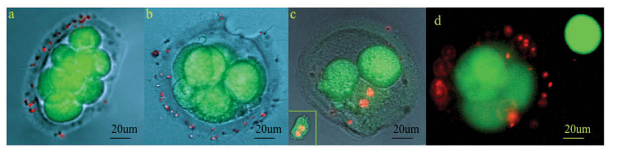
The impact of acidic or laser zona drilling on the viability of human blastocyst cells was assessed using CFSE (green: viable cells) and PI (red: damaged cells).
Figure (a) illustrates an elliptical 8-cell embryo drilled with acidic Tyrode's solution,
Figure (d) is a fluorescence micrograph of a live laser-biopsied embryo and its single blastomere. Observe the dead sperm (red) bound to the embryo's zona pellucida. [2]
The introduction of femtosecond laser technology has not only enhanced the precision and safety of in vivo tissue sampling but also significantly improved the minimally invasive nature and efficiency of surgical procedures. It provides physicians with a means to precisely control the depth and location of incisions, thereby ensuring sample quality while minimizing trauma to the patient. The development and application of this technology signify a shift in medical surgical techniques towards higher precision, increased safety, and greater minimally invasive capabilities, which are of great importance for improving the accuracy of disease diagnosis, reducing surgical risks, and accelerating patient recovery.
In summary, the application of femtosecond laser in biopsy probe drilling technology has not only markedly increased the precision and safety of surgeries but also reduced surgical trauma and patient recovery time, bringing about revolutionary advancements in the medical field. The evolution of this technology has profound implications for enhancing the efficiency and safety of medical surgeries and improving the accuracy of disease diagnosis, heralding a new direction for future medical surgical techniques.
References:
[1] H. Ramakonar et al. "Intraoperative detection of blood vessels with an imaging needle during neurosurgery in humans." Science Advances, 4 (2018). https://doi.org/10.1126/sciadv.aav4992.
[2]K. Chatzimeletiou et al. "Comparison of effects of zona drilling by non-contact infrared laser or acid Tyrode's on the development of human biopsied embryos as revealed by blastomere viability, cytoskeletal analysis and molecular cytogenetics.." Reproductive biomedicine online, 11 6 (2005): 697-710 . https://doi.org/10.1016/S1472-6483(10)61688-4.
Related products